Wax Analysis
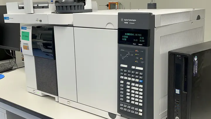 GCMS
GCMSWax analysis
Wax components were analyzed by submerging the freshly peeled discs in 7 mL of chloroform for 30 s twice, and then derivatized by sylation, with BFTSA/pyridine (1:1) at 70°C for 1 h. Samples were injected out of chloroform in a gas chromatograph coupled to a mass spectrometer and a flame ionization detector (Agilent 6890N GC Network systems) on a HP-1 column(30m,0.32mmID,1mm film thickness, (Agilent)) (temperature program: 50°C for 2 min, raise by 40°Cmin–1 to 200°C, held for 2 min at 200°C, raised by 3°C min–1 to 320°C, and held for 30 min at 320°C). Peaks were quantified on the basis of their FID ion current.
Cuticular wax analysis
The cuticular wax composition of leaves and stems of flowering Arabidopsis wild-type (Col-0), lacs1-1, lacs2-3 and lacs1-1 lacs2-3 plants was determined as described by Chen et al. (2003) with slight modifications. After 6 weeks of growth, leaf and stem (cauline leaves and siliques removed) samples were submerged in hexane for 30 sec, the internal standard hexadecane was added and samples were evaporated under nitrogen. Waxes were derivatized by heating at 100°C for 15 min in N,O-bis(trimethylsilyl)trifluoroacetamide (BSTFA). Silylated samples were analyzed by GC with a Hewlett-Packard 5890 series II gas chromatograph (GMI, Inc., http://www.gmi.com) equipped with a flame ionization detector and a 12-m, 0.2-mm HP-1 capillary column with helium as the carrier gas. The column temperature was programmed:an initial temperature of 80°C and increased at 15°C min-1 to 260°C, at which point the temperature remained unchanged for 10 min. The temperature was then increased at 5°C min-1 to 320°C, at which point the temperature was held for 20 min. Quantification was based on flame ionization detector (FID) peak areas relative to the internal standard hexadecane. Specific correction factors were developed from external standards as described by Jenks et al. (1995, 1996). The molecular identities of individual wax compounds were determined by electron impact GC/MS (FinniganMAT/Thermospray Corp., http://www. thermoquest.com).
Analysis of cuticular wax and polyesters
Waxes were extracted from rosette leaves of 4-wk-old Arabidopsis and stems of 6-wk-old Arabidopsis as previously described (Huang et al. 2022). Three grams of 8-wk-old P. patens gametophores were air-dried for 4 h to remove surface moisture and immersed in chloroform for 30 s to extract the waxes. The wax composition was determined as described by Lü et al. (2009). To quantify the amounts of wax components, GC was performed with an Agilent 8860 gas chromatograph equipped with a DB-5 (30 m × 0.25 mm × 0.25 μm, Agilent, CA) capillary column with helium as the carrier gas and a FID. The column temperature was initially set at 80 °C and gradually increased at 40 °C min−1 to 200 °C, followed by an increase to 270 °C at 10 °C min−1; at each time point, the temperature was held for 2 min. The temperature then increased gradually at 3 °C min−1 and finally reached 320 °C, holding for 40 min. Quantification was performed based on FID peak areas relative to the internal standard tetracosane. To analyze cutin polyesters, the leaves of 4-wk-old Arabidopsis plants were analyzed as described by Lü et al. (2009). The total levels of wax and cutin monomers were calculated as the sum of all identified components; unknowns were trace compounds and were not included.
Self
The cuticular wax were extracted from leaves of 4-week-old tomato as previously described (Lü et al., 2009). Tomato leaves immersed in 4 mL of hexane containing tetracosane (25 µg) as an internal standard for 30 s, then the waxes were derivatized by silylation, with BFTSA/pyridine (1:1, v/v) at 70°C for 1 h. Samples were analyzed by GC with an Agilent 7890B GC Network systems (Agilent) equipped with a flame ionization detector and a 122-5532G DB-5MS+DG column (30 m x 0.25 mm x 0.25 µm; Agilent) with helium as the carrier gas. The column temperature program: initial 80 °C held for 2 min, increment of 40 °C/min to 200 °C held for 2 min, 10°C/min up to 270 °C held for 2 min, 3 °C/min up to 320 °C held for 40 min. Quantification was performed based on FID peak areas relative to the internal standard tetracosane.